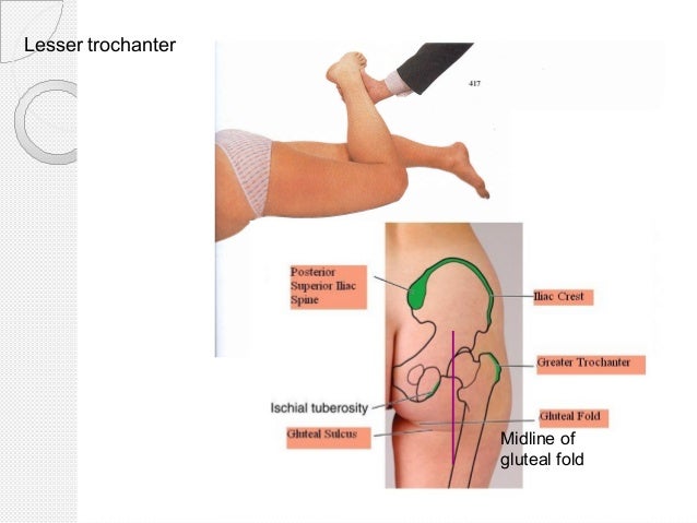
Lesser Trochanter Palpation / Proximal Femur | Team Bone - Palpate for tenderness (± swelling) over the following areas:
Get link
Facebook
X
Pinterest
Email
Other Apps
Lesser Trochanter Palpation / Proximal Femur | Team Bone - Palpate for tenderness (± swelling) over the following areas:. Soleal line of tibia, posterior head of fibula i: The greater trochanter (great trochanter) of the femur is a large, irregular, quadrilateral eminence and a part of the skeletal system. The less common tendinitis and partial tears of the distal biceps tendon present with localized pain and. For the surgeon's orientation, in order to determine the level of the osteotomy, perform a palpation of the lesser trochanter and the fossa piriformis. Small trochanter) is a conical eminence, which varies in size in different subjects;
Repérer et palper le grand trochanter, qui est la principale saillie osseuse de l'aspect latéral de la cuisse proximale. Pii avulsion fractures of the lesser trochanter in adolescents are uncommon. It projects from the lower and back part of the base of the neck. Calcaneus via the achilles tendon palpate. Avulsion fractures of the lesser trochanter in adults are almost always due to metastatic bone disease.
Untitled Document bio.sunyorange.edu from www.bio.sunyorange.edu Pii avulsion fractures of the lesser trochanter in adolescents are uncommon. Calcaneus via the achilles tendon palpate. Isolated lesser trochanter fractures in adults should be considered pathological until proven otherwise. The lesser trochanter (trochanter minor; Study 39 muscle palpation flashcards from lauren s. Bilateral lesser trochanter avulsion fractures in an adolescent. Its principles and the cervical disc. Afin de faciliter le positionnement de la sonde.
The lesser trochanter (trochanter minor;
Bilateral lesser trochanter avulsion fractures in an adolescent. Its principles and the cervical disc. Study 39 muscle palpation flashcards from lauren s. In elderly patients, the most common lesions are metastasis, myeloma or lymphoma. Two muscles insert onto the lesser trochanter With the patient supine, palpate the anterior joint line just lateral to the femoral artery pulsation, below the middle third of the. For the surgeon's orientation, in order to determine the level of the osteotomy, perform a palpation of the lesser trochanter and the fossa piriformis. The less common tendinitis and partial tears of the distal biceps tendon present with localized pain and. The lesser trochanter (trochanter minor; Soleal line of tibia, posterior head of fibula i: Upper extremity of right femur viewed from behind and above. Havept flex hip with your palpation soleus: If you wish to palpate on the skin, provide a private room for assessment.
This injury is a result of a. On this bony prominence attach tendons of the gluteus maximus, medius and. Its principles and the cervical disc. As a long bone, the femur consists of a narrow shaft that is capped at either end. The lesser trochanter is on the medial aspect of the inferior end of the femoral neck.
Tibial tuberosity avulsion fracture | Image | Radiopaedia.org from images.radiopaedia.org The iliopsoas musculotendinous unit can be injured from repetitive, excessive, or unbalanced contraction while. Upper extremity of right femur viewed from behind and above. While the skin can not be viewed with clothes covering the area, the greater trochanter usually can be palpated over clothing. Iliac crest to iliotibial tract. In elderly patients, the most common lesions are metastasis, myeloma or lymphoma. Greater and lesser trochanter fracture causes & treatment. With the patient supine, palpate the anterior joint line just lateral to the femoral artery pulsation, below the middle third of the. The lesser trochanter (small trochanter) of the femur is a conical eminence, which varies in size in different subjects.
We report three patients with avulsion of the lesser trochanter due to neoplasm.
350 x 234 jpeg 11 кб. The iliopsoas musculotendinous unit can be injured from repetitive, excessive, or unbalanced contraction while. Bilateral lesser trochanter avulsion fractures in an adolescent. The greater trochanter is situated on the proximal and lateral side of the femur , just distal to the hip these bursae and the periosteum of the greater trochanter are innervated by a small branch of the. It is directed lateral and medially and slightly posterior. We report three patients with avulsion of the lesser trochanter due to neoplasm. Isolated lesser trochanter fractures in adults should be considered pathological until proven otherwise. Small trochanter) is a conical eminence, which varies in size in different subjects; If you wish to palpate on the skin, provide a private room for assessment. Superior ramus of pubis to just distal of lesser trochanter. A little understood disorder of the vertebral joint. The greater trochanter is located proximally and laterally on the femur, just distal to the hip joint and the neck of the femur. Lesser trochanter bsis are less common compared with femoral neck bsis.
While the skin can not be viewed with clothes covering the area, the greater trochanter usually can be palpated over clothing. On this bony prominence attach tendons of the gluteus maximus, medius and. With the patient supine, palpate the anterior joint line just lateral to the femoral artery pulsation, below the middle third of the. Havept flex hip with your palpation soleus: Study 39 muscle palpation flashcards from lauren s.
Surface anatomy of lower limb from image.slidesharecdn.com On this bony prominence attach tendons of the gluteus maximus, medius and. The iliopsoas musculotendinous unit can be injured from repetitive, excessive, or unbalanced contraction while. Greater and lesser trochanter fracture causes & treatment. It projects from the lower and back part of the base of the neck. Its principles and the cervical disc. Bilateral lesser trochanter avulsion fractures in an adolescent. Superior ramus of pubis to just distal of lesser trochanter. For the surgeon's orientation, in order to determine the level of the osteotomy, perform a palpation of the lesser trochanter and the fossa piriformis.
350 x 234 jpeg 11 кб.
Greater and lesser trochanter fracture causes & treatment. It projects from the lower and back part of the base of the neck. Its principles and the cervical disc. This injury is a result of a. Havept flex hip with your palpation soleus: Isolated lesser trochanter fractures in adults should be considered pathological until proven otherwise. The greater trochanter is situated on the proximal and lateral side of the femur , just distal to the hip these bursae and the periosteum of the greater trochanter are innervated by a small branch of the. The iliopsoas musculotendinous unit can be injured from repetitive, excessive, or unbalanced contraction while. Lesser trochanter is the site of attachment for iliopsoas (forceful contraction of which can cause an avulsion fracture of the lesser. The greater trochanter (great trochanter) of the femur is a large, irregular, quadrilateral eminence and a part of the skeletal system. For the surgeon's orientation, in order to determine the level of the osteotomy, perform a palpation of the lesser trochanter and the fossa piriformis. Small trochanter) is a conical eminence, which varies in size in different subjects; Greater trochanter palpation | gluteal tendinopathy (gtps.
Afin de faciliter le positionnement de la sonde lesser trochanter. In elderly patients, the most common lesions are metastasis, myeloma or lymphoma.


Comments
Post a Comment